

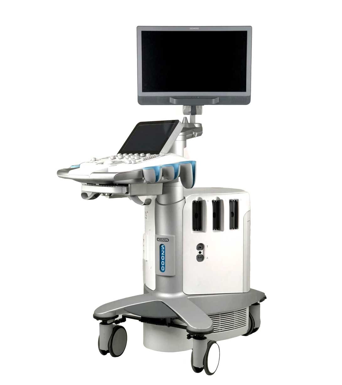
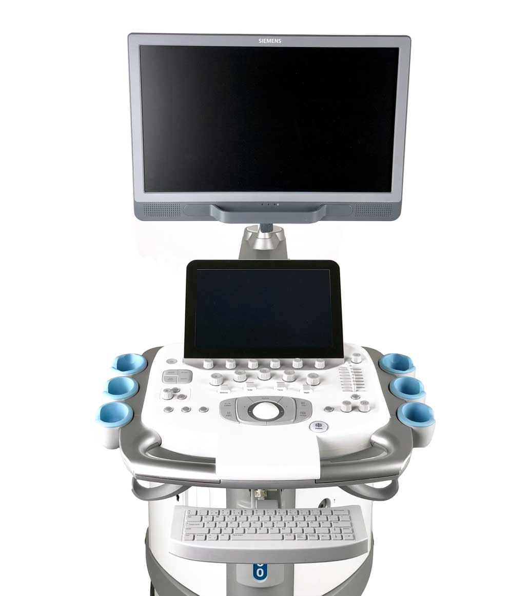

Description
The Siemens S2000 HELX Evolution is a high-precision ultrasound scanner with high-quality images that supports advanced technologies such as PureWave Crystal and eSie Fusion Imaging. The scanner has an ergonomic design with a 19-inch monitor and easy navigation for user convenience. In addition, it has a built-in acoustic sensor to reduce noise and artifacts, which increases diagnostic accuracy. The Siemens S2000 HELX Evolution also has the ability to connect to a DICOM network for data transfer and cloud storage.
The Siemens S2000 HELX Evolution scanner has a variety of scanning modes to perform different types of examinations and ensure maximum diagnostic accuracy:
- 2D mode: used to depict two-dimensional structures and organs.
- 3D mode: provides visualization of three-dimensional objects with the additional ability to manipulate the image.
- 4D mode: provides real-time reproduction of a three-dimensional image, which allows you to observe the movement of objects and detect defects in moving structures.
- Doppler imaging mode: allows you to detect and measure blood flow velocity in blood vessels.
- Color Doppler: allows you to detect blood flow in the form of color signals and images.
- 3D angiography mode: provides three-dimensional reconstruction of blood vessels and visualization of pathologies.
- Elastography: allows to assess tissue stiffness and detect changes associated with pathological processes.
- Specialized imaging modes: allows you to examine various structures and organs, such as mammary glands, cervical vessels, abdominal organs, etc.
The Cardio Package contains a number of additional functions and modes specifically designed for the diagnosis of cardiovascular diseases:
- Heart rate: measures heart rate and records heart cycles.
- Stress ECG: allows you to measure the electrical activity of the heart at rest and during increased physical activity.
Functional echocardiography: allows you to evaluate the functional characteristics of the heart, such as ejection fraction and valve condition. - Percutaneous coronary angiography: allows visualization of coronary arteries and detection of coronary disease.
Stress ECG with image: allows you to measure the electrical activity of the heart at rest and during increased physical activity while simultaneously obtaining a three-dimensional image of the heart.


